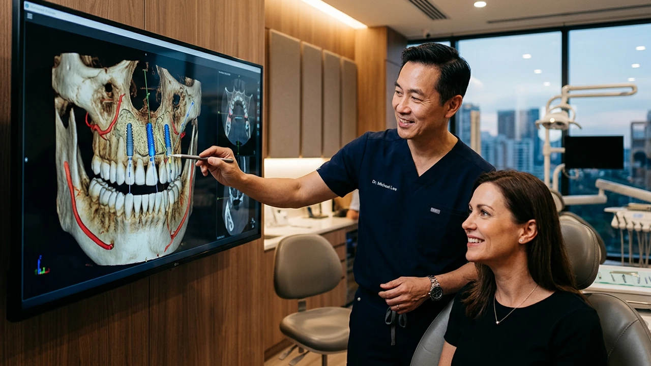
At Ultra Dental Turkey, we assess bone density for dental implants in Turkey through a full clinical review and three-dimensional imaging. We study jawbone height, width, internal structure, gum health, bite force, nearby nerves, and sinus position. This gives every implant a clear role in the final smile.
Reduced bone can still leave several routes open. These include grafting, short implants, tilted implants, All-on-4, pterygoid implants, and zygomatic implants. The right route comes from anatomy, health, healing, and bridge design. This keeps each clinical choice tied to anatomy, comfort, function, travel timing, and long-term care.
Why Bone Density for Dental Implants in Turkey Matters
A dental implant needs a stable bone around it. Bone volume means the height and width available at the implant site. Bone quality describes the outer cortical layer and the inner trabecular pattern. A jaw can have good height and limited width. Another jaw can have enough space and a softer internal pattern. The first grip of an implant is called primary stability. It comes from the contact between the implant and the prepared bone. Over the following weeks, osseointegration develops. During this stage, living bone bonds with the implant surface.
A scan gives us key measurements. Clinical judgement links them with implant design, position, surgical technique, and loading time. This is why jawbone density for dental implants forms one part of a wider assessment.
Cortical Bone and Trabecular Bone
Cortical bone forms the dense outer shell. Trabecular bone forms the inner network. A thicker cortical layer can create a firm entry point. Softer bone may call for careful drilling, added implant support, or a longer healing stage. Very dense bone also needs controlled preparation to manage heat and compression.

What a CBCT Scan Shows Before Implant Surgery
A CBCT scan for dental implants creates cross-sectional and three-dimensional views of the jaw. It reveals areas that a two-dimensional image can hide.
We use it to map:
- Ridge height and width
- Cortical boundaries
- Sinuses, nasal floor, and nerve canals
- Defects, undercuts, retained roots, and pathology
- Implant angles and space for the planned bridge
ITI guidance supports CBCT when cross-sectional imaging adds value after a clinical examination. It also describes CBCT as the imaging choice for three-dimensional implant site assessment. Linear measurements are generally accurate, and a safety margin around vital anatomy remains important.
Bone Height and Width
Height shows the vertical space between the top of the ridge and nearby anatomy. Width shows the space from the cheek side to the tongue or palate side.
These measurements guide implant size and show where grafting, ridge expansion, or angled placement may fit.
Sinuses, Nerves, and Undercuts
In the upper back jaw, the sinus may sit close to the ridge. In the lower jaw, the nerve canal shapes the safe implant zone. Deep undercuts can change the entry angle.
A precise dental implant bone quality assessment keeps surgery linked to real anatomy. We plan around the future crown or bridge from the start.
What CBCT Grey Values Mean
CBCT grey values can show relative differences within one scan. They vary across machines, settings, field size, reconstruction methods, movement, and metal artefacts. This makes them different from universal medical CT density numbers.
For that reason, we read the scan as a full image. We combine grey patterns with cortical thickness, trabecular appearance, bone dimensions, medical history, and findings during surgery.
CBCT Signs That Can Change the Implant Plan
A Narrow Ridge
A narrow ridge may suit guided bone regeneration, ridge expansion, a narrow-diameter implant, or a different implant position.
Reduced Vertical Height
Limited height near the sinus or lower-jaw nerve may lead to short dental implants, sinus elevation, or an angled route. ITI consensus found similar one-to-five-year survival for selected implants measuring 6 mm or less and longer implants in posterior jaws. Long-term care and case selection remain central.
A Low Sinus Floor
The sinus can move closer to the ridge after upper back teeth are lost. This jawbone loss after missing teeth may suit an internal sinus lift when the height change is small. A larger deficit may suit an external sinus lift or a graft-reduced full-arch route.
Thin Cortical Plates
Thin outer bone can raise the need for contour grafting or a staged plan. We also assess the gum tissue, since bone and soft tissue work together around the implant.
Severe Upper-Jaw Resorption
Advanced upper-jaw bone loss may suit pterygoid or zygomatic anchorage. These routes use stronger areas farther back in the jaw or cheekbone. A midface CBCT and sinus review form a key part of zygomatic planning.
Can You Have Dental Implants with Low Bone Density?
Yes. Low bone density dental implants can succeed through careful case selection and a tailored surgical plan.
The D1 to D4 bone quality framework describes bone from very dense to softer internal patterns. It guides discussion rather than acting as a single score.
We also review:
- Jaw location
- Number of missing teeth
- Gum condition
- Bite force
- Teeth grinding
- Smoking
- Diabetes control
- Medicines
- Oral hygiene
- Implant number
- Bridge span
- Cleaning access
For insufficient bone for dental implants, the route may rebuild the ridge or use existing anatomy. We match the implant strategy to the final teeth.
Bone Grafting Choices for Dental Implants in Turkey
A bone graft for dental implants adds or preserves bone at a planned implant site. The technique depends on the shape and size of the defect.
Socket Preservation
Socket preservation places graft material in an extraction site. It aims to reduce ridge shrinkage during healing. This can create a stronger base for a later implant.
Guided Bone Regeneration
Guided bone regeneration uses graft material and a protective membrane. It often suits local width defects or contour gaps around an implant.
Particulate and Block Grafts
Particulate grafts can shape smaller defects. Block grafts can rebuild larger areas. Scan findings, blood supply, soft tissue, and implant position guide the route.
Sinus Lift
A sinus lift for dental implants raises the sinus lining and creates space for graft material in the upper back jaw. The implant may enter during the same visit when stability is strong. A staged route can suit larger defects.
Graft Material Choices
Graft material may come from the patient, screened human tissue, animal-derived material, synthetic material, or a blend. Defect size, volume stability, and surgical technique shape the choice.
NHS guidance states that many grafts heal for three to six months before implant treatment. Larger reconstructions can extend the full timeline.
For UK guests comparing a dental implant bone graft Turkey plan, we explain the graft type, membrane, implant timing, visit count, and final bridge stage in writing.
Fixed-Teeth Alternatives That Can Reduce Major Grafting
Grafting remains valuable. A graft-reduced route may suit some jaws.
Short Implants
Short implants use available vertical bone near the sinus or lower-jaw nerve. They can reduce the scale of vertical augmentation in selected posterior sites. Implant diameter, bite load, crown height, and maintenance all shape the decision.
Narrow-Diameter Implants
Narrow-diameter dental implants can suit selected narrow ridges or tight tooth spaces. The restoration type and chewing load guide their use.
Tilted Implants and All-on-4
All-on-4 with bone loss uses two front implants and two angled rear implants to support a full arch. The angle can use available bone and reduce the need for posterior grafting.
We separate reduced grafting from a universal graft-free promise. A scan can still show a need for local grafting, extra implants, or another route.
For UK guests comparing all on 4 dental implants Turkey, we explain implant distribution, temporary teeth, final bridge material, cleaning access, and healing stages.
All-on-6
All-on-6 uses six implants to spread bridge forces across more support points. It can suit a wider arch, stronger bite, or a bone map with six favourable sites. More implants only add value when anatomy and final bridge design support them.
Pterygoid Implants
Pterygoid implants Turkey treatment uses the back of the upper jaw for added posterior support. It can suit advanced back upper-jaw bone loss and reduce the scale of sinus grafting.
Zygomatic Implants
Zygomatic implants Turkey treatment uses the cheekbone for anchorage in selected cases with severe upper-jaw atrophy, previous implant loss, or earlier graft complications. ITI consensus recognises zygomatic implants as an evidence-based route for major maxillary bone deficiency.
These cases need advanced planning, specialist skill, and clear maintenance.
Grafting or a Graft-Reduced Fixed-Teeth Route?
| Route | Common use | Main strength | Planning point |
|---|---|---|---|
Guided bone regeneration | Local ridge defect | Rebuilds width and contour | Healing and membrane stability |
Sinus lift | Low upper sinus floor | Creates vertical implant space | Sinus health and graft healing |
Short implants | Limited vertical height | Uses existing bone | Bite load and crown height |
All-on-4 | Full-arch tooth loss | Uses angled implant positions | Stability and bridge design |
Pterygoid implants | Back upper-jaw loss | Adds posterior anchorage | Advanced anatomy |
Zygomatic implants | Severe upper-jaw loss | Uses cheekbone support | Specialist surgery and aftercare |
The strongest choice comes from the scan, final teeth, and personal priorities. The table creates a simple starting point for a detailed clinical discussion. It also keeps timing, healing, maintenance, and travel planning clear from the start.
Can Low Bone Density Still Support Same-Day Fixed Teeth?
Immediate loading dental implants can carry a temporary restoration during the first week after implant placement. ITI definitions place immediate loading within that early period. Early loading follows later, and conventional loading starts after a longer healing stage.
A temporary fixed teeth bridge protects appearance and function during healing. The final bridge follows integration, gum healing, bite refinement, and laboratory work.
We assess:
- Implant stability
- Implant spread
- Bone contact
- Bridge rigidity
- Bite pattern
- Grinding
- Cleaning access
- Medical factors
A longer healing route can be the strongest biological strategy for softer bone. Clear timing creates confidence and protects the final result.
Cost, Packages, and the Written Treatment Plan
Dental implants Turkey cost depends on scan findings, implant number, grafting, extractions, sedation, temporary teeth, final bridge material, laboratory work, and review stages.
For implant costs in Turkey, a written plan should list:
- CBCT and examination
- Extractions
- Graft type and membrane
- Implant brand and number
- Temporary restoration
- Final bridge material
- Visit count
- Healing periods
- Medicines
- Transfers or accommodation
- Review route
- Remedial terms
The same detail matters for full mouth dental implants Turkey package deals and full mouth dental implants Turkey price comparisons. Value grows when every clinical stage appears clearly.
At Ultra Dental Turkey, we connect each quote to a scan-led strategy. This keeps dental implants in Antalya focused on health, function, and long-term care. It also gives clear context to Turkey dental packages.
What UK Guests Should Confirm Before Travel
The General Dental Council encourages UK residents to check clinician qualifications, treatment details, aftercare, complaint routes, insurance, and possible remedial costs before overseas care.
Before dental treatment in Turkey, ask for:
- A written diagnosis
- A scan-led implant map
- Implant brand and dimensions
- Graft material details
- Temporary and final tooth stages
- Visit dates
- Recovery guidance
- Aftercare contacts
- Copies of scans and treatment records
We plan travel around surgery and healing, especially where grafting or advanced full-arch surgery forms part of dental implants in Turkey.
How We Plan Low-Bone Cases at Ultra Dental Turkey
We start with your history, photographs, records, and available scans.
We assess bone, gums, bite, facial structure, tooth position, and cleaning access. We then plan the bridge and place implants around that goal.
For fixed teeth with bone loss, we may use standard implants, grafting, angled implants, pterygoid implants, zygomatic implants, or a blended strategy.